How BIS EEG Sensor Technology Measures Anesthesia Depth
What is bispectral index (BIS) monitoring in anesthesia?
Bispectral index (BIS) monitoring translates complex electroencephalogram (EEG) patterns into a dimensionless score (0–100) to quantify anesthesia depth. A BIS value of 40–60 indicates optimal surgical hypnosis, balancing unconsciousness with hemodynamic stability. The technology uses proprietary algorithms to analyze four EEG subparameters:
- Burst suppression ratio (BSR)
- Relative beta ratio
- 95% spectral edge frequency
- Electromyogram (EMG) power
These metrics are weighted differently across anesthesia stages, as demonstrated in a landmark Nature study analyzing 5,427 surgical cases (2019).
From EEG signals to real-time depth assessment: The role of BIS sensors
BIS EEG sensors convert raw brainwave data into actionable insights through three phases:
| Parameter | Function | Dominant Influence Phase |
|---|---|---|
| Burst suppression ratio | Detects isoelectric EEG silence | Deep anesthesia |
| Relative beta ratio | Measures fast vs. slow wave power | Light sedation |
| Spectral edge frequency | Identifies highest active frequency | Transitional anesthesia states |
This multi-parametric approach enables real-time anesthesia adjustments. For example, a sudden EMG power spike (indicating muscle activity) triggers alerts even if BIS scores appear stable.
Technical considerations in signal conduction and sensor performance
Forehead sensor placement and skin-electrode conductivity critically impact data accuracy. Poor adhesion or oily skin increases impedance, artificially inflating BIS values by up to 15 points. Modern sensors integrate artifact-detection algorithms that:
- Filter electrocautery interference
- Compensate for signal drift
- Auto-validate EEG quality every 6 seconds
Clinical validation studies show median absolute errors of ≤4.1 BIS points compared to manual EEG interpretation (BMC Anesthesiology, 2018). However, manufacturers disclose only 60% of algorithmic logic, requiring careful clinical correlation.
The Science Behind BIS: Bispectral Analysis and EEG Signal Processing
Understanding bispectral analysis for anesthesia depth evaluation
Bispectral analysis takes those raw EEG signals and turns them into numbers we can actually work with by looking at how different frequencies relate to each other in phase. The technique started out back in seismic research and oceanography before finding its way into medicine. What makes it special is that it picks up on these complex patterns in brain waves that seem to match up with how anesthesia affects patients. Traditional methods just look at how strong the signal is and what frequencies are present. But bispectral approaches go further by spotting when different waves interact with one another. This matters a lot because it helps tell the difference between someone who's lightly sedated around BIS 60 to 80 range versus fully anesthetized in the lower 40 to 60 range.
Processing raw EEG into a reliable BIS value: Algorithms and validation
The BIS EEG sensors look at several key factors when assessing brain activity. These include things like burst suppression ratio (BSR), electromyogram power readings, spectral edge frequency measurements, plus what they call the relative beta ratio. According to research published back in 2019, these sensors actually use machine learning algorithms that crunch all this data through some pretty complex math formulas known as weighted regression equations. The results? An average error range of about plus or minus 4.1 BIS units. They tested this system on nearly 5,500 different surgeries and found it works reasonably well regardless of who the patient is or what kind of anesthesia they get. Still worth noting though, most companies aren't telling us everything about how their algorithms work since around 30% remains under wraps as proprietary information. This lack of complete openness definitely raises questions about just how transparent we really are being with patients and medical staff.
How the BIS score correlates with brain activity under anesthesia
Studies indicate that Bispectral Index (BIS) readings tend to match up with what's happening in the cortex when it comes to suppression, but they don't really reflect what's going on deeper in the brain structures. Take sevoflurane for example at 1 MAC dose level, this typically brings down BIS scores to around 32 as it quiets those thalamocortical connections. Interesting thing though, ketamine does the opposite effect. Even though patients are clearly dissociated under ketamine, their BIS numbers actually go up to about 59 instead. A validation study back in 2018 confirmed these strange differences between drugs. This explains why BIS monitoring devices essentially measure how sedatives affect brain waves rather than giving us some kind of definitive measure of actual anesthetic depth during surgery.
Proper BIS Sensor Placement and Non-Invasive EEG Acquisition
Optimal Forehead Placement for Accurate BIS EEG Sensor Readings
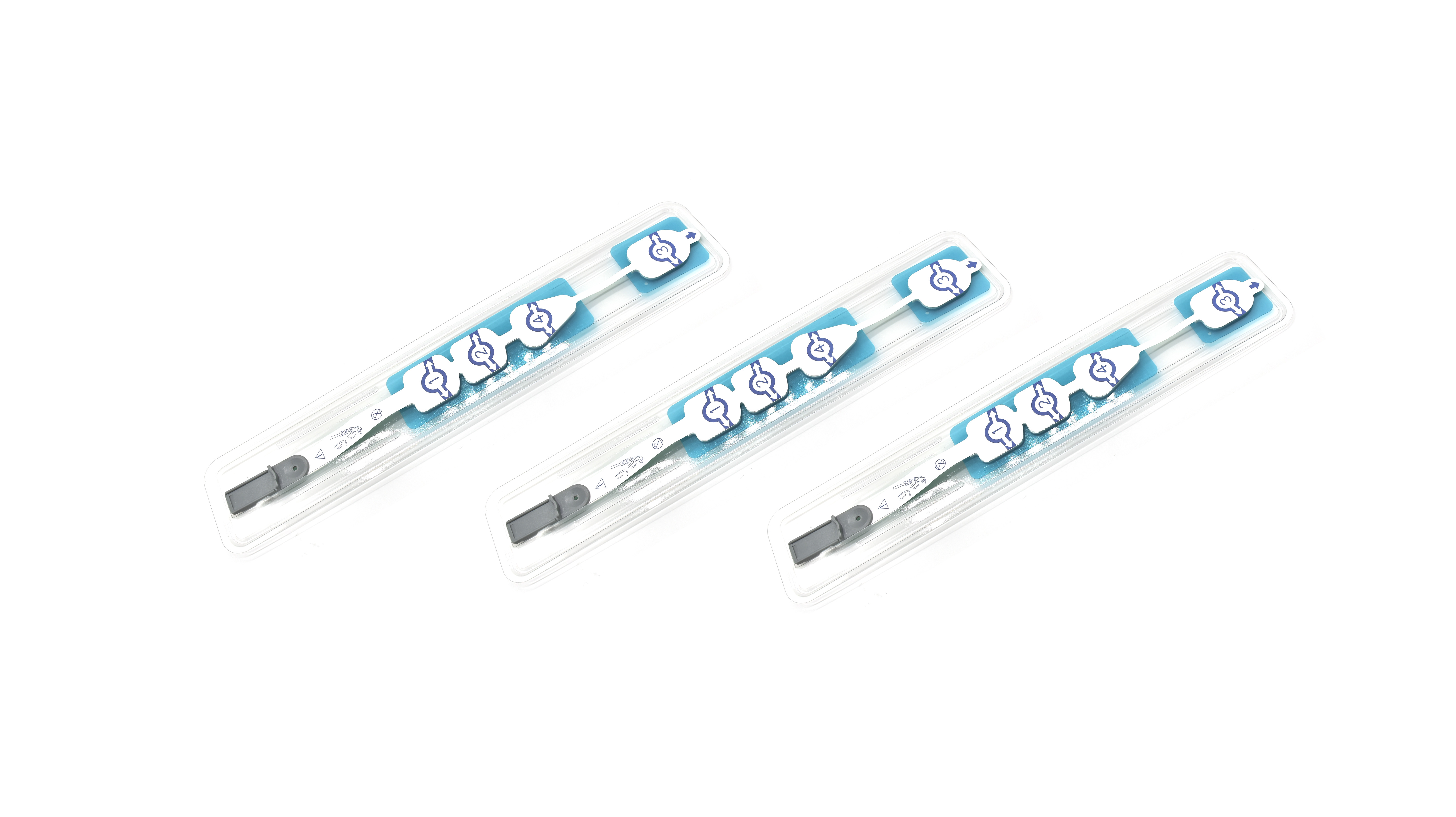
BIS EEG sensors achieve optimal signal capture through standardized forehead placement. Research shows positioning electrodes diagonally across the forehead and temple aligns with frontotemporal brain activity patterns critical for anesthesia monitoring. This placement minimizes muscle interference while maintaining consistent contact with superficial cerebral electrical activity.
Using Skin Electrodes for Reliable, Non-Invasive Anesthesia Monitoring
The latest BIS systems now feature these super thin hydrogel electrodes which cut down on skin resistance around 40 percent when compared to old fashioned adhesive options. The sensors are medically certified and can stay attached to patients for anywhere between twelve to twenty four hours without causing any discomfort. This makes them really useful during long surgical procedures where brain activity monitoring needs to be ongoing. When doctors properly prepare the skin first cleaning it with alcohol wipes and getting rid of extra hair they've seen about a third improvement in signal quality according to recent studies in hospitals across the country.
Minimizing Artifacts and Ensuring Signal Quality During Surgery
Three key strategies prevent intraoperative signal distortion:
- Isolating EEG leads from electrosurgical devices (>30 cm separation)
- Using shielded cables to reduce electromagnetic interference by 55%
- Implementing adaptive filters that suppress high-frequency artifacts like EMG (>30 Hz)
Studies confirm these measures reduce false BIS readings by 74% during cautery use or patient movement. Regular impedance checks (<5 kΩ) further validate sensor functionality mid-procedure.
Interpreting BIS Values in Clinical Practice
BIS Scale Explained: Sedation, Unconsciousness, and Anesthetic Endpoints
The bispectral index (BIS) quantifies anesthesia depth on a 0–100 scale, with lower values indicating deeper suppression of brain activity. Clinical guidelines categorize consciousness states as follows:
- 60–100: Light sedation to fully awake
- 40–60: General anesthesia (optimal surgical range)
- <40: Deep hypnotic state (risk of burst suppression)
The proprietary BIS algorithm weighs four EEG parameters—burst suppression ratio (BSR), electromyogram (EMG) power, spectral edge frequency (SEF), and relative beta ratio (RBR)—differently across five BIS ranges. This explains why sudden EMG spikes (>65 dB) can falsely elevate scores by 20+ points despite adequate anesthesia.
Linking EEG Patterns to Levels of Consciousness During Anesthesia
BIS sensors work by turning those raw EEG signals we get from patients into actual information doctors can use. They do this by looking at specific patterns that tell us about how different drugs are affecting the brain. Take propofol for instance. This drug tends to knock down those fast beta waves between around 13 and 30 Hz, which makes the RBR numbers go up as someone gets more sleepy. When someone is under surgery level anesthesia with BIS readings between 40 and 60, their SEF usually settles somewhere around 15 to 18 Hz. But watch out for those burst suppression periods where over half of the brain activity stops completely. That's when things get too deep. Some interesting research has shown that ketamine actually raises BIS scores even though patients aren't conscious at all. This shows just how varied these brain wave responses can be depending on what medication is being used.
Limitations of BIS: Why It Doesn’t Predict Movement or Hemodynamic Response
BIS does a good job tracking what's happening in the cortex, but falls short when it comes to those deeper brain pathways that control things like reflex movements and blood pressure shifts. Looking back at a study from 2018, around one in five patients had BIS readings below 40 yet still moved during surgery, which shows there are definitely gaps in how well it picks up on spinal cord activity. Another issue is that sometimes the body has strong reactions to pain signals without any noticeable change in BIS levels. This means doctors need other ways to monitor these responses, such as checking heart rate variability, to get the full picture of what's going on inside the patient.
Clinical Validation and Reliability of BIS EEG Sensors in Surgery
Development and clinical trials behind the BIS monitor
To check if the BIS monitoring system actually works, researchers ran multiple rounds of clinical trials with around 1,500 participants. They wanted to see how brain wave patterns matched up with what doctors could observe during sedation. One big study across several medical centers showed up in Frontiers in Medicine. The results were pretty impressive really. When using BIS guidance for anesthesia, there was an 82% drop in patients becoming aware during surgery among those 2,463 risky operations. Based on all this evidence, most experts now agree that keeping BIS scores between 40 and 60 is best practice for proper anesthesia depth. The company behind BIS also tweaked their software algorithms so they can better ignore muscle activity signals that might otherwise confuse readings.
Evidence from surgical case studies on BIS-guided anesthesia
Looking at data from 36 controlled trials involving around 7,761 patients shows that brain function monitoring (BIS) cuts down the chance of patients waking up during surgery by about 35%, which is pretty significant compared to just watching standard vital signs. But wait there's another side to this story. The B-Unaware study found similar results when they compared BIS monitoring against measuring breath gases at the end of exhalation for certain types of anesthesia, so it really depends on what kind of procedure we're talking about here. For neurological operations specifically, these brain sensors seem to make a real difference. They help reduce problems with thinking after surgery in roughly 23% of cases because doctors can adjust sedatives much more accurately than before.
Adoption trends and trust in BIS technology across medical centers
More than 85 percent of academic hospitals have started using BIS sensors as part of their regular anesthesia procedures according to recent surveys from 2023. Why? Well, these devices have been around for over two decades now, and throughout that time there's been very few issues reported with them - something like less than one percent of problems actually related to the equipment itself. There are still some tricky situations though when patients experience extreme hypothermia or have pacemakers installed. But don't get me wrong, most of the time things work just fine. A study published in Nature back in 2020 found that BIS remains accurate about 94 times out of 100 during normal surgery conditions. That kind of track record explains why so many medical facilities continue relying on this technology for their perioperative care needs.
FAQ
What is BIS monitoring and how does it work?
BIS (Bispectral Index) monitoring uses EEG patterns to assess anesthesia depth with a score from 0 to 100. A BIS value between 40 and 60 is ideal for surgical hypnosis, combining unconsciousness and hemodynamic stability. It uses algorithms to analyze subparameters such as burst suppression ratio, beta ratio, spectral edge frequency, and electromyogram power.
What are BIS sensors and how are they placed?
BIS sensors are placed on the forehead and temple to capture frontotemporal brain activity patterns. They are used for accurate brain monitoring during anesthesia.
Why don't BIS scores reflect movement or hemodynamic response?
BIS captures cortical activity but doesn't adequately measure deeper brain pathways that govern reflex movements and blood pressure changes, making it insufficient for monitoring spinal cord activity.
Table of Contents
- How BIS EEG Sensor Technology Measures Anesthesia Depth
- The Science Behind BIS: Bispectral Analysis and EEG Signal Processing
- Proper BIS Sensor Placement and Non-Invasive EEG Acquisition
- Optimal Forehead Placement for Accurate BIS EEG Sensor Readings
- Using Skin Electrodes for Reliable, Non-Invasive Anesthesia Monitoring
- Minimizing Artifacts and Ensuring Signal Quality During Surgery
- Interpreting BIS Values in Clinical Practice
- Clinical Validation and Reliability of BIS EEG Sensors in Surgery
- FAQ